Publications
2024
-
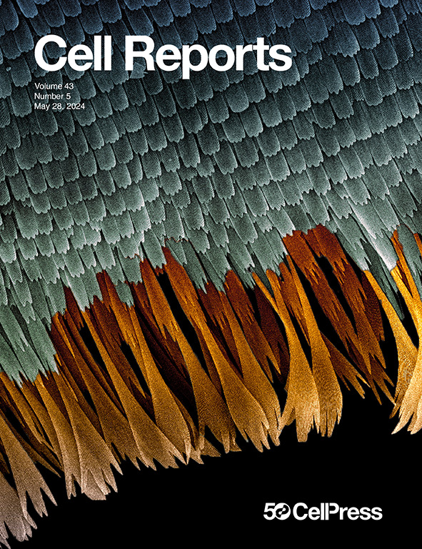
 The Janus kinase 1 is critical for pancreatic cancer initiation and progressionHridaya Shrestha, Patrick D Rädler, Rayane Dennaoui, and 8 more authorsCell Reports, 2024
Interleukin-6 (IL-6)-class inflammatory cytokines signal through the Janus tyrosine kinase (JAK)/signal transducer and activator of transcription (STAT) pathway and promote the development of pancreatic ductal adenocarcinoma (PDAC); however, the functions of specific intracellular signaling mediators in this process are less well defined. Using a ligand-controlled and pancreas-specific knockout in adult mice, we demonstrate in this study that JAK1 deficiency prevents the formation of KRASG12D-induced pancreatic tumors, and we establish that JAK1 is essential for the constitutive activation of STAT3, whose activation is a prominent characteristic of PDAC. We identify CCAAT/enhancer binding protein δ (C/EBPδ) as a biologically relevant downstream target of JAK1 signaling, which is upregulated in human PDAC. Reinstating the expression of C/EBPδ was sufficient to restore the growth of JAK1-deficient cancer cells as tumorspheres and in xenografted mice. Collectively, the findings of this study suggest that JAK1 executes important functions of inflammatory cytokines through C/EBPδ and may serve as a molecular target for PDAC prevention and treatment.
2021
- Scientific ReportsDual recombinase action in the normal and neoplastic mammary gland epitheliumPatrick D Rädler, Kerry Vistisen, Aleata A Triplett, and 7 more authorsScientific Reports, 2021
We developed a transgenic mouse line that expresses the codon-optimized Flp recombinase under the control of the MMTV promoter in luminal epithelial cells of the mammary gland. In this report, we demonstrate the versatile applicability of the new MMTV-Flp strain to manipulate genes in a temporally and spatially controlled manner in the normal mammary gland, in luminal-type mammary tumors that overexpress ERBB2, and in a new KRAS-associated mammary cancer model. Although the MMTV-Flp is expressed in a mosaic pattern in the luminal epithelium, the Flp-mediated activation of a mutant KrasG12D allele resulted in basal-like mammary tumors that progressively acquired mesenchymal features. Besides its applicability as a tool for gene activation and cell lineage tracing to validate the cellular origin of primary and metastatic tumor cells, we employed the MMTV-Flp transgene together with the tamoxifen-inducible Cre recombinase to demonstrate that the combinatorial action of both recombinases can be used to delete or to activate genes in established tumors. In a proof-of-principle experiment, we conditionally deleted the JAK1 tyrosine kinase in KRAS-transformed mammary cancer cells using the dual recombinase approach and found that lack of JAK1 was sufficient to block the constitutive activation of STAT3. The collective results from the various lines of investigation showed that it is, in principle, feasible to manipulate genes in a ligand-controlled manner in neoplastic mammary epithelial cells, even when cancer cells acquire a state of cellular plasticity that may no longer support the expression of the MMTV-Flp transgene.
- Cancer Metastasis R...Models of pancreatic ductal adenocarcinomaRayane Dennaoui, Hridaya Shrestha, and Kay-Uwe WagnerCancer and Metastasis Reviews, 2021
Although pancreatic cancer remains to be a leading cause of cancer-related deaths in many industrialized countries, there have been major advances in research over the past two decades that provided a detailed insight into the molecular and developmental processes that govern the genesis of this highly malignant tumor type. There is a continuous need for the development and analysis of preclinical and genetically engineered pancreatic cancer models to study the biological significance of new molecular targets that are identified using various genome-wide approaches and to better understand the mechanisms by which they contribute to pancreatic cancer onset and progression. Following an introduction into the etiology of pancreatic cancer, the molecular subtypes, and key signaling pathways, this review provides an overview of the broad spectrum of models for pancreatic cancer research. In addition to conventional and patient-derived xenografting, this review highlights major milestones in the development of chemical carcinogen-induced and genetically engineered animal models to study pancreatic cancer. Particular emphasis was placed on selected research findings of ligand-controlled tumor models and current efforts to develop genetically engineered strains to gain insight into the biological functions of genes at defined developmental stages during cancer initiation and metastatic progression.
-
 Highly metastatic claudin-low mammary cancers can originate from luminal epithelial cellsPatrick D Rädler, Barbara L Wehde, Aleata A Triplett, and 7 more authorsNature communications, 2021
Claudin-low breast cancer represents an aggressive molecular subtype that is comprised of mostly triple-negative mammary tumor cells that possess stem cell-like and mesenchymal features. Little is known about the cellular origin and oncogenic drivers that promote claudin-low breast cancer. In this study, we show that persistent oncogenic RAS signaling causes highly metastatic triple-negative mammary tumors in mice. More importantly, the activation of endogenous mutant KRAS and expression of exogenous KRAS specifically in luminal epithelial cells in a continuous and differentiation stage-independent manner induces preneoplastic lesions that evolve into basal-like and claudin-low mammary cancers. Further investigations demonstrate that the continuous signaling of oncogenic RAS, as well as regulators of EMT, play a crucial role in the cellular plasticity and maintenance of the mesenchymal and stem cell characteristics of claudin-low mammary cancer cells.
2020
- Scientific ReportsEfficient tissue-type specific expression of target genes in a tetracycline-controlled manner from the ubiquitously active Eef1a1 locusKazuhito Sakamoto, Patrick D Rädler, Barbara L Wehde, and 8 more authorsScientific Reports, 2020
Using an efficient gene targeting approach, we developed a novel mouse line that expresses the tetracycline-controlled transactivator (tTA) from the constitutively active Eef1a1 locus in a Cre recombinase-inducible manner. The temporally and spatially controlled expression of the EF1-LSL-tTA knockin and activation of tTA-driven responder transgenes was tested using four transgenic lines that express Cre under tissue-specific promoters of the pancreas, mammary gland and other secretory tissues, as well as an interferon-inducible promoter. In all models, the endogenous Eef1a1 promoter facilitated a cell-type-specific activation of target genes at high levels without exogenous enhancer elements. The applicability of the EF1-LSL-tTA strain for biological experiments was tested in two studies related to mammary gland development and tumorigenesis. First, we validated the crucial role of active STAT5 as a survival factor for functionally differentiated epithelial cells by expressing a hyperactive STAT5 mutant in the mammary gland during postlactational remodeling. In a second experiment, we assessed the ability of the EF1-tTA to initiate tumor formation through upregulation of mutant KRAS. The collective results show that the EF1-LSL-tTA knockin line is a versatile genetic tool that can be applied to constitutively express transgenes in specific cell types to examine their biological functions at defined developmental stages.
2018
-
 Janus Kinase 1 Plays a Critical Role in Mammary Cancer ProgressionBarbara L Wehde, Patrick D Rädler, Hridaya Shrestha, and 3 more authorsCell Reports, 2018
Janus kinases (JAKs) and their downstream STAT proteins play key roles in cytokine signaling, tissue homeostasis, and cancer development. Using a breast cancer model that conditionally lacks Janus kinase 1, we show here that JAK1 is essential for IL-6-class inflammatory cytokine signaling and plays a critical role in metastatic cancer progression. JAK1 is indispensable for the oncogenic activation of STAT1, STAT3, and STAT6 in ERBB2-expressing cancer cells, suggesting that ERBB2 receptor tyrosine kinase complexes do not directly activate these STAT proteins in vivo. A genome-wide gene expression analysis revealed that JAK1 signaling has pleiotropic effects on several pathways associated with cancer progression. We established that FOS and MAP3K8 are targets of JAK1/STAT3 signaling, which promotes tumorsphere formation and cell migration. The results highlight the significance of JAK1 as a rational therapeutic target to block IL-6-class cytokines, which are master regulators of cancer-associated inflammation.
- Molecules and Cellsδ-Catenin increases the stability of EGFR by decreasing c-Cbl interaction and enhances EGFR/Erk1/2 signaling in prostate cancerNensi Shrestha*, Hridaya Shrestha*, Taeyong Ryu, and 8 more authorsMolecules and cells, 2018
δ-Catenin, a member of the p120-catenin subfamily of armadillo proteins, reportedly increases during the late stage of prostate cancer. Our previous study demonstrates that δ-catenin increases the stability of EGFR in prostate cancer cell lines. However, the molecular mechanism behind δ-catenin-mediated enhanced stability of EGFR was not explored. In this study, we hypothesized that δ-catenin enhances the protein stability of EGFR by inhibiting its lysosomal degradation that is mediated by c-casitas b-lineage lymphoma (c-Cbl), a RING domain E3 ligase. c-Cbl monoubiquitinates EGFR and thus facilitates its internalization, followed by lysosomal degradation. We observed that δ-catenin plays a key role in EGFR stability and downstream signaling. δ-Catenin competes with c-Cbl for EGFR binding, which results in a reduction of binding between c-Cbl and EGFR and thus decreases the ubiquitination of EGFR. This in turn increases the expression of membrane bound EGFR and enhances EGFR/Erk1/2 signaling. Our findings add a new perspective on the role of δ-catenin in enhancing EGFR/Erk1/2 signaling-mediated prostate cancer.
2017
- Cellular SignallingHakai, an E3-ligase for E-cadherin, stabilizes δ-catenin through Src kinaseHridaya Shrestha, Taeyong Ryu, Young-Woo Seo, and 8 more authorsCellular Signalling, 2017
Hakai ubiquitinates and induces endocytosis of the E-cadherin complex; thus, modulating cell adhesion and regulating development of the epithelial-mesenchymal transition of metastasis. Our previous published data show that δ-catenin promotes E-cadherin processing and thereby activates β-catenin-mediated oncogenic signals. Although several published data show the interactions between δ-catenin and E-cadherin and between Hakai and E-cadherin separately, we found no published report on the relationship between δ-catenin and Hakai. In this report, we show Hakai stabilizes δ-catenin regardless of its E3 ligase activity. We show that Hakai and Src increase the stability of δ-catenin synergistically. Hakai stabilizes Src and Src, which in turn, inhibits binding between glycogen synthase kinase-3β and δ-catenin, resulting in less proteosomal degradation of δ-catenin. These results suggest that stabilization of δ-catenin by Hakai is dependent on Src.
2016
- BBA MCRInvestigation of the molecular mechanism of δ-catenin ubiquitination: Implication of β-TrCP-1 as a potential E3 ligaseHridaya Shrestha*, Tingting Yuan*, Yongfeng He, and 8 more authorsBiochimica et Biophysica Acta (BBA)-Molecular Cell Research, 2016
Ubiquitination, a post-translational modification, involves the covalent attachment of ubiquitin to the target protein. The ubiquitin-proteasome pathway and the endosome-lysosome pathway control the degradation of the majority of eukaryotic proteins. Our previous study illustrated that δ-catenin ubiquitination occurs in a glycogen synthase kinase-3 (GSK-3) phosphorylation-dependent manner. However, the molecular mechanism of δ-catenin ubiquitination is still unknown. Here, we show that the lysine residues required for ubiquitination are located mainly in the C-terminal portion of δ-catenin. In addition, we provide evidence that β-TrCP-1 interacts with δ-catenin and functions as an E3 ligase, mediating δ-catenin ubiquitin-proteasome degradation. Furthermore, we prove that both the ubiquitin-proteasome pathway and the lysosome degradation pathway are involved in δ-catenin degradation. Our novel findings on the mechanism of δ-catenin ubiquitination will add a new perspective to δ-catenin degradation and the effects of δ-catenin on E-cadherin involved in epithelial cell-cell adhesion, which is implicated in prostate cancer progression.
- Scientific ReportsInteraction of EGFR to δ-catenin leads to δ-catenin phosphorylation and enhances EGFR signalingYongfeng He, Taeyong Ryu, Nensi Shrestha, and 7 more authorsScientific Reports, 2016
Expression of δ-catenin reportedly increases during late stage prostate cancer. Furthermore, it has been demonstrated that expression of EGFR is enhanced in hormone refractory prostate cancer. In this study, we investigated the possible correlation between EGFR and δ-catenin in prostate cancer cells. We found that EGFR interacted with δ-catenin and the interaction decreased in the presence of EGF. We also demonstrated that, on one hand, EGFR phosphorylated δ-catenin in a Src independent manner in the presence of EGF and on the other hand, δ-catenin enhanced protein stability of EGFR and strengthened the EGFR/Erk1/2 signaling pathway. Our findings added a new perspective to the interaction of EGFR to the E-cadherin complex. They also provided novel insights to the roles of δ-catenin in prostate cancer cells.


